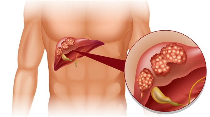


TA cyst is the medical term used to describe a space of roundish or saclike shape in some part of the body. It may be empty or contain watery or mucus types of fluid. It is not uncommon to find one or several small cysts in the liver when a patient has an ultrasound scan or CAT scan of the abdomen for some reason. The vast majority of these cysts are found by chance as they do not produce any symptoms.
These "simple cysts" usually arise because a small area of liver cells die or degenerate. The most common cause is advancing years and poor diet and lifestyle. Sometimes these cysts can be full of "fatty material" in those with a fatty liver. These cysts do not represent liver disease because the liver, being such a huge organ, has plenty of other areas containing healthy cells to enable liver function to remain normal.
If your doctor finds that you have one or several of these degenerative types of liver cysts (simple cysts), do not become alarmed. Obviously you do not want to develop many more of these cysts and thankfully it is easy to stop them from multiplying. In many cases it is possible to gradually shrink the cysts using nutritional medicine.
The cause of simple liver cysts is not known, but they are believed to be congenital in origin. The cysts are lined by biliary-type epithelium and perhaps result from progressive dilatation of biliary microhamartomas (are small nodules in or near portal tracts). These cysts contain a fluid which has an electrolyte composition mimicking plasma. The cyst fluid is continually secreted by the epithelial lining of the cyst. Simple cysts generally cause no symptoms. However, symptoms such as
A dull right upper quadrant pain is common if the cyst is large in size.
Patients may also report abdominal bloating and feeling full.
Occasionally, a palpable abdominal mass is possible.
Jaundice caused by bile duct obstruction is rare,
Patients with mobile cysts that twist around (torsion) is rare but can be presented with an acute pain abdomen.
Liver tumors (cystadenomas and cystadenocarcinoma) with central necrosis visualized on imaging studies are often misdiagnosed as liver cysts. True intrahepatic neoplastic cysts are rare. The cause of cystadenomas and cystadenocarcinomas is unknown, but they may represent the growth and development of abnormal embryonic analogs of the gallbladder or biliary epithelium. These cystic tumors are lined with biliary-type cuboidal or columnar cells and are surrounded by ovarianlike stroma. Cystadenoma is a premalignant lesion with neoplastic transformation to cystadenocarcinoma confirmed by tubulopapillary architecture and invasion of the basement membrane.Cystadenoma most often occurs in middle-aged women. However, cystadenocarcinoma equally affects both men and women. Most patients are asymptomatic or have vague abdominal complaints of bloating, nausea, and fullness. These patients, like all those with hepatic cysts, eventually present with abdominal pain. Rarely, they present with evidence of biliary obstruction.
Hydatid cysts are caused by infestation with the parasite Echinococcus granulosus. This parasite is found worldwide, but it is particularly common in areas of sheep and cattle farming. The adult tapeworm lives in the digestive tract of carnivores, such as dogs or wolves. Eggs are released into the stool and are inadvertently ingested by the intermediate hosts, such as sheep, cattle, or humans. The egg larvae invade the bowel wall and mesenteric vessels of the intermediate host, allowing circulation to the liver. In the liver, the larvae grow and become encysted. The hydatid cyst develops an outer layer of inflammatory tissue and an inner germinal membrane that produces daughter cysts. When carnivores ingest the liver of the intermediate host, the scolices of the daughter cysts are released in the small intestines and grow into adult worms, thus completing the life cycle of the worm.
The evaluation of a patient with a simple liver cyst involves carefully recording patient history and performing a physical examination plus an imaging study, such as an abdominal CT scan, to define the anatomy of the cyst. It becomes important to distinguish a cyst from a benign tumor or an abscess as treatment will vary for each condition
Treatment of PCLD or solitary nonparasitic cysts of the liver is indicated in symptomatic patients. Asymptomatic patients do not require therapy because the risk of developing complications related to the lesion is lower than the risk associated with treatment.
Patients with hydatid cysts should be treated to prevent complications related to cyst growth and rupture. If cysts on imaging studies show abnormalities suggestive of cystic tumors, resection is indicated. Abscesses should be treated at the time of identification, but percutaneous drainage and antibiotics are usually adequate treatment.