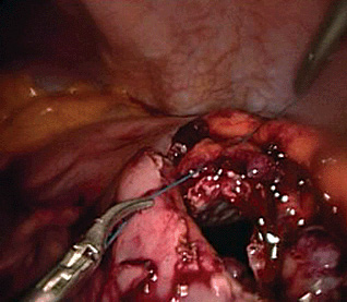

Pancreatic pseudocyst was first described by Morgan. It is a collection of fluid rich in pancreatic enzymes, necrotic tissue and blood usually located at the lesser sac as a result of a pancreatic inflammatory process. Its lining is made of non-epithelialised granulation tissue and therefore the name pseudocyst (pseudo - false) . Contrary to this, a true cyst should have an epithelial lining. It mainly occurs as a complication of acute pancreatitis but may also occur after abdominal trauma, as a complication of an acute exacerbation or progressive ductal obstruction in chronic pancreatitis and rarely due to gallbladder disease . About 75% of all pancreatic masses are pancreatic pseudocysts .
Pancreatic pseudocysts may occur within or outside the pancreas, solitary or multiple and may be small or large. Most patients may be asymptomatic . The disease may mimic other medical conditions and therefore needs a careful clinical evaluation. Symptoms may include abdominal pain, nausea and vomiting, fever, abdominal mass/distension and weight loss among others.
Diagnosis can be achieved through a careful clinical and radiological evaluation especially abdominal ultrasonography and/or CT scan.
The pancreas produces fluid rich in digestive enzymes. Acute pancreatitis leads to extravasation of pancreatic secretions rich in enzymes that result to digestion of adjoining tissues. This leads to disruption of pancreatic parenchyma and surrounding tissues. Collection of fluid containing pancreatic enzymes, heamolysed blood and necrotic debri occurs around the pancreas mainly in the lesser sac since it is a potential space. The collection/s may resolve spontaneously as the patient recovers from the acute phase but others may become organized and become walled-off by a thick wall of granulation tissue and fibrosis over several weeks forming a pancreatic pseudocyst .
It is important to exclude all other differential diagnosis before subjecting the patient to surgery. A complete medical history and physical exam is mandatory. Diagnostic procedures for pancreatic pseudocyst may include;
Blood tests. Laboratory studies may be limited.
Abdominal Ultrasonography. Repeated ultrasonography can be used to monitor the progression of pancreatitis at various stages.
Computed Tomography Scan (CT scan). CT scan has been the standard test with 90%-100% sensitivity . It gives information regarding fluid collection as well as presence or absence of necrosis.
Magnetic Resonance Imaging (MRI). MRI helps to differentiate between organized necrotic tissue and pseudocyst as well as accessing both the biliary and pancreatic ductal systems in selected patients.
Endoscopic Ultrasonography (EUS). Useful in endoscopic drainage.
Endoscopic Retrograde Cholangiopancreatography(ERCP). This can be performed selectively on patients suspected to have ampullary obstruction, all recurrent pseudocysts and patients who develop pseudocysts long after acute recovery of acute episode of pancreatitis.
